
Glaucoma testing.
Eye pressure is one part of a larger picture. Glaucoma assessment considers your optic nerves, side vision, corneas and health history, often before you notice a change in sight.
Understanding your risk.
Glaucoma damages the optic nerve. The most common form often develops slowly, with few early symptoms. Risk assessment helps an eye doctor decide which tests and follow-up are appropriate.
Your eye doctor will ask about age, family history, ancestry, previous eye conditions, injury or surgery, and steroid medicines. Risk patterns differ between types of glaucoma, including among people of African or Black Caribbean, Hispanic or Asian ancestry. Eye pressure, corneal thickness, optic-nerve appearance, nearsightedness and farsightedness also help inform the assessment. Farsightedness can be associated with narrower drainage angles; nearsightedness is associated with greater open-angle glaucoma risk.
Tell your clinician about diabetes, blood-pressure problems, migraines, circulation concerns and smoking history. These are parts of your overall health history, not proof that you have glaucoma.
Angle-closure glaucoma can develop suddenly. Intense eye pain, a red eye, nausea or sudden blurred vision need urgent medical attention.
Measuring eye pressure.
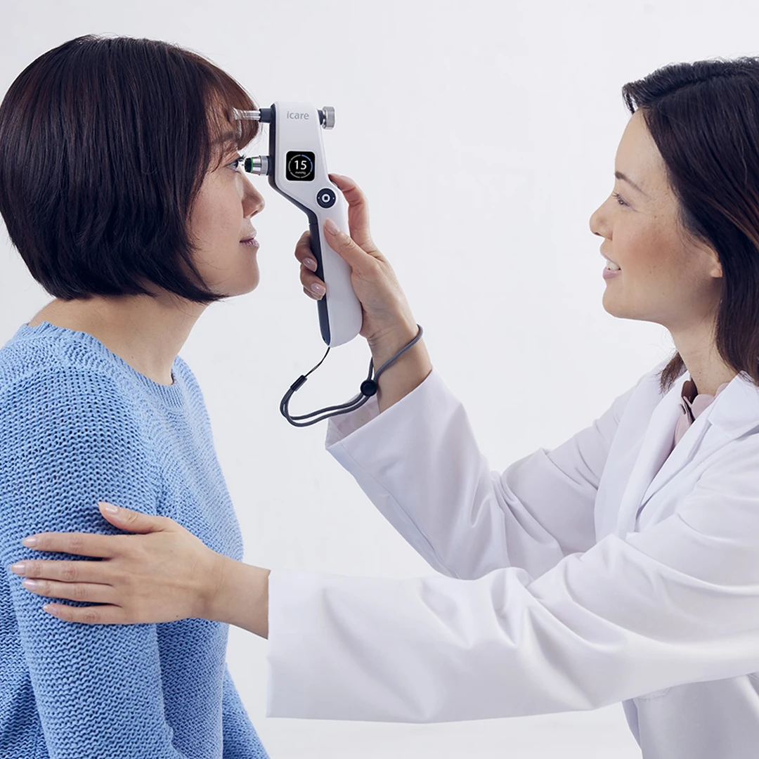
Tonometry measures the pressure inside the eye. iDoctor uses iCare IC100 rebound measurement, which does not require an air puff or anesthetic drops.
A pressure reading alone cannot diagnose or rule out glaucoma. Some people have high pressure without glaucoma, while others develop glaucoma at pressures considered normal. Your eye doctor interprets the number alongside the other findings.
Looking at the optic nerve.
Ophthalmoscopy examines the shape and appearance of the optic nerve. Dilating drops may be used to widen the pupil and provide a better view. The examination can guide whether more testing is needed.
Other tests your doctor may consider.
Perimetry.
A visual field test maps how well you see in different areas, including your side vision. You look toward a target and respond when you notice lights. Testing may be repeated to check reliability and monitor change. Follow-up frequency depends on your risk and findings.
Gonioscopy.
Gonioscopy examines the drainage angle where the iris and cornea meet. After numbing drops, a special contact lens with mirrors is placed on the eye so the clinician can inspect whether the angle is open or narrow.
Pachymetry.
Pachymetry measures the thickness of the cornea. Corneal thickness can influence pressure readings and helps your clinician interpret the overall assessment.
Follow-up matters.
Glaucoma diagnosis is based on the combined findings, not one test or a single pressure cutoff. Ask your eye doctor which tests you need and when to return.